Optical monitoring of blood coagulability during cardiovascular surgery via coherence-gated DLS
Light scattering-based approaches have proved robust for monitoring the structural dynamics of complex media thanks to their capability to retrieve collective dynamic information through the intensity fluctuations of the light scattered. In my talk I will address the specific features of fiber-based implementations of optical sensing techniques based on spatio-temporal coherence-gated dynamic light scattering (DLS). This sensing approach has a number of unique capabilities such as an effective isolation of single scattering, a large sensitivity and high collection efficiency, and it can also operate over a wide range of optical regimes while providing means for proper ensemble averaging. I will present the application of this sensing technique in a scenario that falls beyond the capabilities of traditional light scattering-based techniques: the real-time monitoring of blood coagulability during cardiovascular surgeries.
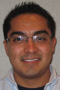
Rafael Guzman is currently a PhD student in Dr. Dogariu’s research group. He received his B.Sc. from the University of Guanajuato, Mexico in 2010, and his M.Sc. from the University of Tamaulipas, Mexico in 2013. He also received a M.Sc. from the University of Central Florida in 2016. His research focuses on the development of fiber-based implementations of optical sensing techniques based on spatio-temporal coherence-gated DLS, for the measurement of the structural and micro-rheological properties of dynamic complex media. His work relies on the interdepartmental cooperation at UCF, industry collaborations with Malvern, partnership with Arnold Palmer Hospital for Children, as well as collaborations with Mexican educational institutions in projects funded by CONACyT. Scientific achievements in his multidisciplinary research (optics, materials science, and biomedical) include one book chapter, over 25 peer-reviewed journal publications, some of which have been published in the top journals of the different areas such as Nature Biomedical Engineering, over 30 conference proceedings, and over 150 citations, in different areas including digital processing of medical images, fiber optics devices and sensors, and optical sensing of dynamics in colloids, non-ergodic media, and biological fluids.
Runners-up

Deep Learning Cell Imaging through Anderson Localizing Optical Fiber
We demonstrate a deep-learning-based fiber imaging system which can transfer real-time artifact-free cell images through a meter-long Anderson localizing optical fiber. The cell samples are illuminated by an incoherent LED light source. A deep convolutional neural network is applied to the image reconstruction process. The network training uses data generated by a set-up with straight fiber at room temperature (~20 °C) but can be utilized directly for high fidelity reconstruction of cell images that are transported through fiber with a few degrees bend and/or fiber with segments heated up to 50 °C. In addition, cell images located several millimeters away from the bare fiber end can be transported and recovered successfully without the assistance of any distal optics. We further evidence that the trained neural network is able to reconstruct the images of cells which are never used in the training process and feature very different morphology.
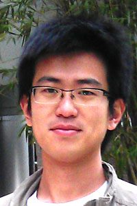
Jian Zhao is currently a Ph.D. candidate in Dr. Axel Schülzgen’s Fiber Optics group. He received the B.S. degree in optics from the Sun Yat-sen University in 2012, and M.S. degree in Optics from CREOL at the University of Central Florida in 2014. His current primary research interest lies in designing, fabricating and application of micro-structured optical fiber as well as deep learning application in optical fiber imaging. Additional research interests include optical frequency comb and high-power femtosecond laser system for high harmonic generation.

Accelerating x-ray diffraction tomography: challenges and solutions
X-ray computed tomography (CT) captures the three-dimensional (3D) interior of the sample in medical diagnosis and industrial inspections. Conventional CT relies on the attenuation of x-ray beam to produce contrast, which is often low among different soft tissues, liquids or low-density polymers. X-ray diffraction tomography (XDT) combines the diffracted x-ray as a structural probe with tomographic imaging to distinguish materials with similar electron densities. However, due to the intrinsically weak diffraction signal, XDT scan using a table-top x-ray source typically takes tens of hours to image the whole 3D volume. The long imaging time and the consequent high radiation exposure pose challenges to the integration of x-ray diffraction imaging in practical applications. In this talk, I will discuss efforts to accelerate x-ray diffraction tomography via designing novel illumination and image acquisition schemes with high collection efficiency, as well as data processing techniques tailored to reconstruction from low photon-flux measurement.
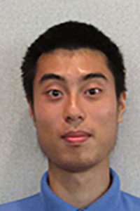
Zheyuan Zhu is currently a PhD student in Optical Imaging System Lab led by Dr. Pang. He received the B.S. degree in physics from Nanjing University in China. His research focuses on designing and prototyping computational imaging platforms with superior performance in contrast, resolution and sensitivity using minimal system resources in both visible and x-ray regimes. He has authored and coauthored 9 journal papers and delivered 4 oral presentations at SPIE conferences. He is also an active member in educational outreach events both within and outside CREOL.